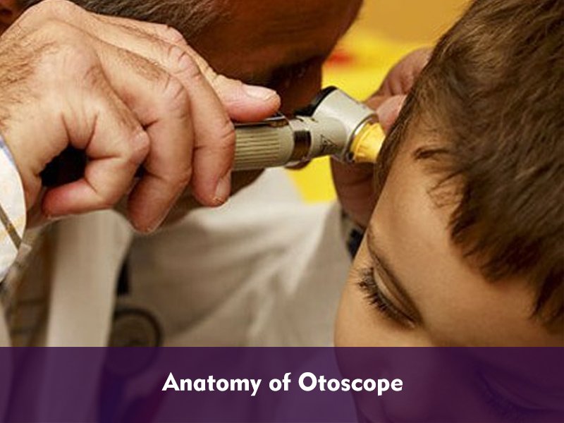
An otoscope, also known as auriscope is a medical instrument used to look inside the ears. The main purpose of this device is to identify any ear infections or ailments of the ear.
With the help of an otoscope, it is possible to clearly view the canal, drum, and tympanic membrane of the ear. Since the eardrum is the differentiating line between the middle ear and external ear, hence, any issue in it can be an indication of ailments in the ear. Infections in the eardrum can be caused due to the shedding of the skin, skin edema, deposit of ear wax or cerumen, and numerous ear ailments. In order to identify and combat all such ailments, you must understand the different parts of an otoscope and how is it used.
An otoscope consists of various parts. In order to use it properly, it is essential to have a complete understanding of different parts of an otoscope. This can be known clearly through the anatomy of an otoscope.
Related : Best Ear Endoscope
Anatomy of an Otoscope
Let’s explore the in-depth anatomy or parts of an otoscope.
- Eartips or Specula
The front or distal end of an otoscope holds a disposable plastic attachment known as specula. The specual allows safe visualization of distinct parts of the inner ear. The specula are available in various lengths and diameters, basically ranging from adults to kids. An otoscope for kids will have small sized specula, while one designed for adults have specula with a bigger diameter.
- Otoscope Head
The next part of an otoscope is its head. The specula are attached to this part. The head of an otoscope is useful to examine the nose, throat, and ear. The light or illumination feature is provided to the otoscope head in order to render clear visibility.
Premium otoscope models incorporate fiberoptic illumination to deliver an obstruction-free visual experience. The head can be fitted with Xenon, LED, or Halogen lights. An entry-level otoscope model usually incorporates a vacuum lamp.
- Magnifying Glass
With the help of a magnifying glass, users are able to get 3 to 4 times better visualization inside the ear. Most of such glasses swivel out of the way to enable instrumentation. Premium otoscope models have magnifying glasses, which can be completely removed.
- Insufflator Port
An insufflator port is useful to perform pneumatic otoscopy. It allows connection of an insufflator bulb to the device.
- Power Switch
The power switch of an otoscope can be located at different places of the device, based on the model. Typically, pocket-sized otoscope has a simple off/on power switch.
The full-size otoscope models come with a rheostatic power switch that allows regulation of light intensity.
- Battery Handle
The battery handle may either consist of rechargeable batteries or may need an external source of power. This part is used to charge the battery of the device and supply the power to various other parts for uninterrupted functionality.
- Battery Cap
The bottom part of an otoscope is known as battery cap. You can unscrew this part in order to conveniently change the battery of your device.
How to Use an Otoscope
Based on the kind of model, otoscopes are available in pocket and standard sizes, perfect to use in a plethora of distinct environments where portability is important. Bigger otoscope models have a chrome-plated, tough brass exterior and come with a reheostatic power switch to enable you to adjust the brightness of the device during the examination.
Before you begin the examination process, it is of utmost importance to select an otoscope with an accurately sized speculum. This is important for convenient examination and maintaining patient’s comfort. You need to select smaller sized specula to examine younger patients with a narrow ear canal. Wide sized specula are ideal to examine adult patients.
Now, when you begin the examination process, it is essential to manipulate the various angles of a patient’s head. This will let you clearly visualize the various structures in the ear.
Next, hold your otoscope in the left hand when assessing the left ear and in right hand when analyzing the right ear. This will serve you the most comfortable access when analyzing the patient’s ear. It is slightly difficult to achieve an ideal angle of view in infants and younger patients. Hence, you need to be extra careful when manipulating their ear.
When you insert the speculum into the ear canal of the patient, it is necessary to be alert and take care so that you don’t cause any damage to the ear’s surrounding tissues. Though the speculum is designed in a way that it does not easily reach the eardrum, however, it can cause discomfort or scratch if not angled properly.
Patients met with ear infections may experience some pain or discomfort during the examination process. But if there is too much resistance then you must stop the examination process at once. Otherwise, it could cause an injury in the ear.
Always use the magnifying glass, lights, and other parts of an otoscope skillfully. When used properly, your otoscope will be an indispensable device for you!

Dr. David Taylor is a medical professional and an avid blogger. He holds an M.D. from Drexel University & a Ph.D. from Indiana University School of Medicine.
Dr. David loves to utilize technology to improve healthcare and he does it daily through BestRatedDocs.com. He founded the company in 2016 with the vision to make the discoverability of the best healthcare facilities & best products simple and easy. His passion for informatics and using technology to empower healthcare professionals and the patients they serve is unmatched. He regularly blogs about technology, health IT, medical products and other healthcare topics at bestrateddocs.com.